

Pinned Items
Recent Activities
-
 Post is under moderationStream item published successfully. Item will now be visible on your stream.
 -
Types of Dental X-rays
There are two main types of dental x-rays: intraoral (the x-ray film is inside the mouth) and extraoral (the x-ray film is outside the mouth).
Intraoral x-rays are the most common type of x-ray. There are several types of intraoral x-rays. Each shows different aspects of teeth.
Bite-wing x-rays show details of the upper and lower teeth in one area of the mouth. Each bite-wing shows a tooth from its crown (the exposed surface) to the level of the supporting bone. Bite-wing x-rays detect decay between teeth and changes in the thickness of bone caused by gum disease. Bite wing x-rays can also help determine the proper fit of a crown (a cap that completely encircles a tooth) or other restorations (eg, bridges). It can also see any wear or breakdown of dental fillings.
Periapical x-rays show the whole tooth — from the crown, to beyond the root where the tooth attaches into the jaw. Each periapical x-ray shows all teeth in one portion of either the upper or lower jaw. Periapical x-rays detect any unusual changes in the root and surrounding bone structures.
Occlusal x-rays track the development and placement of an entire arch of teeth in either the upper or lower jaw.
Extraoral x-rays are used to detect dental problems in the jaw and skull. There are several types of extraoral x-rays.
Panoramic x-rays show the entire mouth area — all the teeth in both the upper and lower jaws — on a single x-ray. This x-ray detects the position of fully emerged as well as emerging teeth, can see impacted teeth, and help diagnosis tumors.
Tomograms show a particular layer or “slice” of the mouth and blur out other layers. This x-ray examines structures that are difficult to clearly see because other nearby structures are blocking the view.
Cephalometric projections show an entire side of the head. This x-ray looks at the teeth in relation to the jaw and profile of the individual. Orthodontists use this x-ray to develop each patient’s specific teeth realignment approach.
Another test that uses x-rays is called a sialogram. This test uses a dye, which is injected into the salivary glands so they can be seen on x-ray film (Salivary glands are a soft tissue that would not be seen with an x-ray.) Dentists might order this test to look for salivary gland problems, such as blockages, or Sjogren’s syndrome (a disorder with symptoms including dry mouth and eyes; this disorder can play a role in tooth decay).
Dental computed tomography (CT) is a type of imaging that looks at interior structures in 3-D (three dimensions). This type of imaging is used to find problems in the bones of the face such as cysts, tumors, and fractures.
Cone Beam CT is a type of x-ray that creates 3-D images of dental structures, soft tissue, nerves, and bone. It helps guide tooth implant placement and evaluates cysts and tumors in the mouth and face. It also can detect problems in the gums, roots of teeth, and jaws. Cone beam CT is similar to regular dental CT in some ways. They both produce accurate and high quality images. However, the way images are taken is different. The cone-beam CT machine rotates around the patient’s head, capturing all data in one single rotation. The traditional CT scan collects “flat slices” as the machine makes several revolutions around the patient’s head. This method also exposes patients to higher level of radiation. A unique advantage of cone beam CT is that it can be used in a dentist’s office. Dental computed CT equipment is only available in hospitals or imaging centers.
Digital imaging is a 2-D type of dental imaging that allows images to be sent directly to a computer. The images can be viewed on screen, stored, or printed out in a matter of seconds. Digital imaging has several other advantages compared with traditional x-rays. The image taken of a tooth, for example, can be enhanced and enlarged. This makes it easier for your dentist to see the tiniest changes that can’t be seen in an oral exam. Also, if necessary, images can be sent electronically to another dentist or specialist for a second opinion or to a new dentist (eg, if you move). Digital imaging also uses less radiation than x-rays.
MRI imaging is an imaging method that takes a 3-D view of the oral cavity including jaw and teeth. (This is ideal for soft tissue evaluation.)Post is under moderationStream item published successfully. Item will now be visible on your stream. -
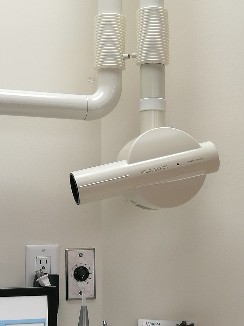
Dental X-ray unit installed in a dental office
 Post is under moderationStream item published successfully. Item will now be visible on your stream.
 -
Why dental X-rays are performed:
Dental X-rays are typically performed yearly. They can happen more often if your dentist is tracking the progress of a dental problem or treatment.
Factors affecting how often you get dental X-rays may include:
your age
your current oral health
any symptoms of oral disease
a history of gum disease (gingivitis) or tooth decay
If you’re a new patient, you’ll probably undergo dental X-rays so that your new dentist can get a clear picture of your dental health. This is especially important if you don’t have any X-rays from your previous dentist.
Children may need to have dental X-rays more often than adults because their dentists might need to monitor the growth of their adult teeth. This is important because it can help the dentist determine if baby teeth need to be pulled to prevent complications, such as adult teeth growing in behind baby teeth.
Risks of dental X-rays
While dental X-rays do involve radiation, the exposed levels are so low that they’re considered safe for children and adults. If your dentist uses digital X-rays instead of developing them on film, your risks from radiation exposure are even lower.
Your dentist will also place a lead “bib” over your chest, abdomen, and pelvic region to prevent any unnecessary radiation exposure to your vital organs. A thyroid collar may be used in the case of thyroid conditions. Children and women of childbearing age may also wear them along with the lead bib.
Pregnancy is an exception to the rule. Women who are pregnant or believe they may be pregnant should avoid all types of X-rays. Tell your dentist if you believe you are pregnant, because radiation is not considered safe for developing fetuses.
Preparing for dental X-rays
Dental X-rays require no special preparation. The only thing you’ll want to do is brush your teeth before your appointment. That creates a more hygienic environment for those working inside your mouth. X-rays are usually done before cleanings.
At the dentist’s office, you’ll sit in a chair with a lead vest across your chest and lap. The X-ray machine is positioned alongside your head to record images of your mouth. Some dental practices have a separate room for X-rays, while others perform them in the same room as cleanings and other procedures.Post is under moderationStream item published successfully. Item will now be visible on your stream. -
Dental X-rays (radiographs) are images of your teeth that your dentist uses to evaluate your oral health. These X-rays are used with low levels of radiation to capture images of the interior of your teeth and gums. This can help your dentist to identify problems, like cavities, tooth decay, and impacted teeth.
Dental X-rays may seem complex, but they’re actually very common tools that are just as important as your teeth cleanings.Post is under moderationStream item published successfully. Item will now be visible on your stream.
There are no activities here yet